Retina
Definition
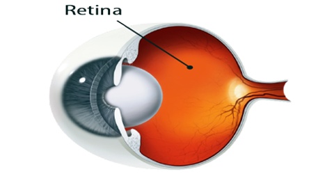
Retina is the light-sensitive membrane that lines the inside of the back of the eyeball and connects to the brain by the optic nerve. The retina of vertebrate animals contains rods and cones, specialized cells that absorb light. Neural retina typically refers to three layers of neural cells (photo receptor cells, bipolar cells, and ganglion cells) within the retina, while the entire retina refers to these three layers plus a layer of pigmented epithelial cells.
It is located near the optic nerve. The purpose of the retina is to receive light that the lens has focused, convert the light into neural signals, and send these signals on to the brain for visual recognition.
Retina is a layered structure with several layers of neurons interconnected by synapses. The only neurons that are directly sensitive to light are the photoreceptor cells. For vision, these are of two types: the rods and cones. Rods function mainly in dim light and provide black-and-white vision while cones support the perception of colour. A third type of photoreceptor, the photosensitive ganglion cells, is important for entrainment and reflexive responses to the brightness of light.
Structure and Functions of Retina
Retina is a complex transparent tissue consisting of several layers, only one of which contains light-sensitive photoreceptor cells. Light must pass through the overlying layers to reach the photoreceptor cells, which are of two types, rods and cones that are differentiated structurally by their distinctive shapes and functionally by their sensitivity to different kinds of light. Basically, the retina processes a picture from the focused light, and the brain is left to decide what the picture is.
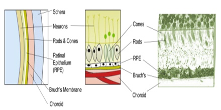
The outer layer contains nerve cells called ganglions contain axons that transmit optic messages to the brain. This layer of nerve cells and the main optic nerve they connect to are actually brain tissue and are considered part of the central nervous system. Pigmented layer is also called retinal epithelium or RPE. These cells are a single layer of hexagonal cells of pigment bits tightly packed together. The important pigment of these cells is what protects the retina from the damaging effects of sunlight.
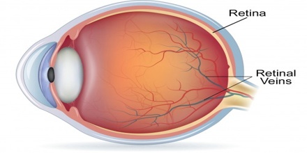
The choroid layer contains connective tissue with many blood vessels. It delivers nourishment and oxygen to the outer layers of the retina. It consists of Haller’s layer, with larger blood vessels, Sattler’s layer, with medium sized veins and arteries, and choriocapillaries, a layer of very fine capillaries.
Retina is located at the back of the eye and this part of the eye contains multiple layers of sensory tissue. The retina has millions of photosensitive cells known as photoreceptors which work to capture and convert light rays into electric signals. Photoreceptors are categorized in two types and these are the rods and cones.
Rods predominate in nocturnal animals and are most sensitive to reduced light intensities; in humans they provide night vision and aid in visual orientation. Cones are more prominent in humans and those animals that are active during the day and provide detailed vision and colour perception. In general, the more cones per unit area of retina, the finer the detail that can be discriminated by that area. Rods are fairly well distributed over the entire retina, but cones tend to concentrate at two sites: the fovea centralis, a pit at the rear of the retina, which contains no rods and has the densest concentration of cones in the eye, and the surrounding macula lutea, a circular patch of yellow-pigmented tissue about 5 to 6 mm (0.2 to 0.24 inch) in diameter.
Reference: healthline.com, britannica.com, naturaleyecare.com, wikipedia.