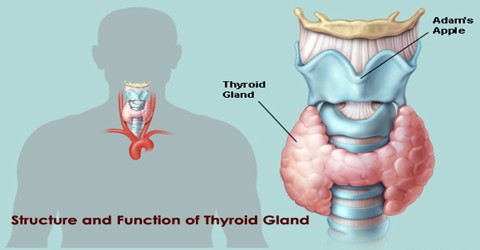
Structure and Function of Thyroid Gland
Introduction
Thyroid Gland is a two-lobed gland that wraps around the trachea and is located at the base of the neck in vertebrate animals. The thyroid gland secretes two important hormones: thyroxine, which regulates the cell metabolism necessary for normal growth and development, and calcitonin, which stimulates the formation of bone and helps regulate the amount of calcium in the blood.
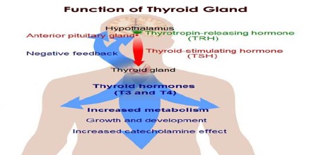
Hormonal output from the thyroid is regulated by thyroid-stimulating hormone (TSH) secreted from the anterior pituitary gland, which itself is regulated by thyrotropin-releasing hormone (TRH) produced by the hypothalamus.
The thyroid gland or thyroid is a highly vascular, brownish-red gland located anteriorly in the lower neck, extending from the level of the fifth cervical vertebra down to the first thoracic. The gland varies from an H to a U shape and is formed by 2 elongated lateral lobes with superior and inferior poles connected by a median isthmus, with an average height of 12-15 mm, overlying the second to fourth tracheal rings.
Structure of Thyroid Gland
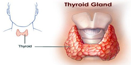
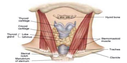
The thyroid is a butterfly-shaped gland that sits low on the front of the neck. The thyroid has two side lobes, connected by a bridge (isthmus) in the middle. When the thyroid is its normal size, we can’t feel it. The thyroid weighs 25 grams in adults, with each lobe being about 5 cm long, 3 cm wide and 2 cm thick, and the isthmus about 1.25 cm in height and width. The gland is usually larger in women, and increases in size in pregnancy.
Principal innervation of the thyroid gland derives from the autonomic nervous system. Parasympathetic fibers come from the vagus nerves, and sympathetic fibers are distributed from the superior, middle, and inferior ganglia of the sympathetic trunk. These small nerves enter the gland along with the blood vessels. Autonomic nervous regulation of the glandular secretion is not clearly understood, but most of the effect is postulated to be on blood vessels, hence the perfusion rates of the glands.
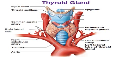
The thyroid sits near the front of the neck, lying against and around the front of the larynx and trachea. The thyroid cartilage and cricoid cartilage lie just above the gland, below the Adam’s apple. The thyroid gland is covered by a thin fibrous capsule, which has an inner and an outer layer. The outer layer is continuous with the pretracheal fascia, attaching the gland to the cricoid and thyroid cartilages, via a thickening of the fascia to form the posterior suspensory ligament of thyroid gland also known as Berry’s ligament. This causes the thyroid to move up and down with swallowing. The inner layer extrudes into the gland and forms the septae that divides the thyroid tissue into microscopic lobules. Typically four parathyroid glands, two on each side, lie on each side between the two layers of the capsule, at the back of the thyroid lobes.
The lobules are composed of follicles, the structural units of the gland, which consist of a layer of simple epithelium enclosing a colloid-filled cavity.
Epithelial cells are of 2 types: principal cells (ie, follicular) and parafollicular cells (ie, C, clear, light cells). Principal cells are responsible for formation of the colloid (iodothyroglobulin), whereas parafollicular cells produce the hormone calcitonin, a protein central to calcium homeostasis. Parafollicular cells lie adjacent to the follicles within the basal lamina.
The thyroid is supplied with arterial blood from the superior thyroid artery, a branch of the external carotid artery, and the inferior thyroid artery, a branch of the thyrocervical trunk, and sometimes by an anatomical variant the thyroid ima artery, which has a variable origin.
Functions of Thyroid Gland
The primary function of the thyroid gland or thyroid is the production of the iodine-containing thyroid hormones, triiodothyronine (T3) and thyroxine (T4) and the peptide hormone calcitonin. T3 is so named because it contains three atoms of iodine per molecule and T4 contains four atoms of iodine per molecule. The thyroid hormones have a wide range of effects on the human body. The thyroid’s hormones regulate vital body functions, including:
- Breathing
- Heart rate
- Central and peripheral nervous systems
- Body weight
- Muscle strength
- Menstrual cycles
- Body temperature
- Cholesterol levels
The thyroid has two sides called lobes that lie on either side of your windpipe, and is usually connected by a strip of thyroid tissue known as an isthmus. Some people do not have an isthmus, and instead have two separate thyroid lobes.
The thyroid is part of the endocrine system, which is made up of glands that produce, store, and release hormones into the bloodstream so the hormones can reach the body’s cells. The thyroid gland uses iodine from the foods you eat to make two main hormones: Triiodothyronine (T3), Thyroxine (T4).
It is important that T3 and T4 levels are neither too high nor too low. Two glands in the brain—the hypothalamus and the pituitary communicate to maintain T3 and T4 balance.