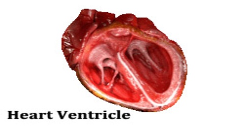
Heart Ventricle
Definition
Ventricle is a chamber of the heart that receives blood from one or more atria and pumps it by muscular contraction into the arteries. Mammals, birds, and reptiles have two ventricles; amphibians and fish have one. The right ventricle receives blood from the right atrium and pumps it into the lungs via the pulmonary artery, and the left ventricle receives blood from the left atrium and pumps it into the circulation system via the aorta.
The heart is divided into four chambers that are connected by heart valves. The lower two chambers of the heart are called heart ventricles. A ventricle is a cavity or chamber that can be filled with fluid, such as the cerebral ventricles. The heart ventricles are separated by a septum into the left ventricle and the right ventricle. The upper two heart chambers are called atria. Atria receive blood returning to the heart from the body and ventricles pump blood from the heart to the body.
Structure and Functions of Ventricle
The left ventricle is one of four chambers of the heart. It is located in the bottom left portion of the heart below the left atrium, separated by the mitral valve. The left ventricle is the thickest of the heart’s chambers and is responsible for pumping oxygenated blood to tissues all over the body. The most common is left ventricular hypertrophy, which causes enlargement and hardening of the muscle tissue that makes up the wall of the left ventricle, usually as a result of uncontrolled high blood pressure.
Left ventricle receives blood from the left atrium and pumps it to the aorta. Blood returning to the heart from the lungs enters the left atrium and passes through the mitral valve to the left ventricle. Blood in the left ventricle is then pumped to the aorta as the ventricles contract and the aortic valve opens. The aorta carries and distributes oxygen-rich blood to the rest of the body.
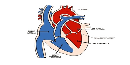
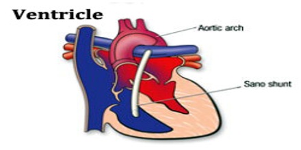
The right ventricle is one of the heart’s four chambers. It is located in the lower right portion of the heart below the right atrium and opposite the left ventricle. It is equal in size to that of the left ventricle, and contains roughly 85 millilitres (3 imp fl oz; 3 US fl oz) in the adult. Its upper front surface is circled and convex, and forms much of the sternocostal surface of the heart. It’s under surface is flattened, forming part of the diaphragmatic surface of the heart that rests upon the diaphragm.
Right ventricle receives blood from the right atrium and pumps it to the main pulmonary artery. Blood passes from the right atrium through the tricuspid valve into the right ventricle. Blood is then forced into the main pulmonary artery as the ventricles contract and pulmonary valve opens. The pulmonary artery extends from the right ventricle and branches into left and right pulmonary arteries. These arteries extend to the lungs.
Clinical Significance
Cardiac conduction is the rate at which the heart conducts the electrical impulses that drive the cardiac cycle. Heart failure is a condition that is caused by the failure of heart ventricles to pump blood efficiently. Cardiac dysrhythmia is an irregular heartbeat that can occur in the ventricles or atria. Normally the heartbeat is initiated in the SA node of the atrium but initiation can also occur in the Purkinje fibres of the ventricles giving rise to premature ventricular contractions, also called ventricular extra beats.
Ventricular tachycardia is another disorder of the heart ventricles. In ventricular tachycardia, the heartbeat is accelerated but the heartbeats are regular. Ventricular tachycardia may lead to ventricular fibrillation, a condition in which the heart beats both rapidly and irregularly. Ventricular fibrillation is the primary cause of sudden cardiac death as the heart beats so quickly and irregularly that it becomes unable to pump blood.
Reference: thoughtco.com, medicinenet.com, healthline.com, wikipedia.