Olfactory Bulb
Definition
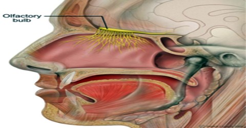
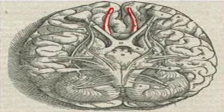
Olfactory bulb is the anterior and slightly enlarged end of the olfactory tract, from which the cranial nerves concerned with the sense of smell originate. It is a key part of the olfactory apparatus consisting of a bulbous enlargement of the end of the olfactory nerve on the under surface of the frontal lobe of each cerebral hemisphere just above the nasal cavity and also called Morgagni’s tubercle.
It receives signals from the olfactory receptor neurons in the nasal epithelium via the olfactory nerves and sends those to the olfactory cortex. In the olfactory bulb various layers can be distinguished. The axons of the olfactory neurons enter the outer layer of the olfactory bulb, the (olfactory) nerve fiber layer, organized into fascicles. The olfactory axons terminate in the glomerular layer, where they synapse with the dendrites of mitral cells, forming the spherical olfactory glomeruli.
Olfactory bulb comprise of granule cell which are a type of interneuron. These granule cells are devoid of axons and hence can create inhibition at local level. This is accomplished by granule cells by giving out GABA from the dedicated spines of dendritic which are in contact with secondary and horizontally long dendrites related to mitral men do not have access to view this node.
Structure and Function of Olfactory Bulb
The Olfactory bulb performs the important task of controlling the nerves related to odor in the brain system. It is the most rostral (forward) part of the brain, as seen in rats. In humans, however, the olfactory bulb is on the inferior (bottom) side of the brain. The axons of olfactory receptor (smell receptor) cells extend directly into the highly organized olfactory bulb, where information about odours is processed.
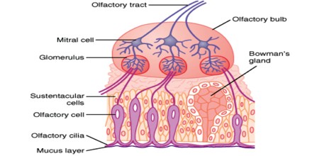
Within the olfactory bulb are discrete spheres of nerve tissue called glomeruli. They are formed from the branching ends of axons of receptor cells and from the outer (dendritic) branches of interneurons, known in vertebrates as mitral cells, that pass information to other parts of the brain. Tufted cells, which are similar to but smaller than mitral cells, and periglomerular cells, another type of interneuron cell, also contribute to the formation of glomeruli.
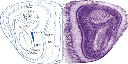
The main olfactory bulb has a multi-layered cellular architecture. In order from surface to the center the layers are:
- Glomerular layer
- External plexiform layer
- Mitral cell layer
- Internal plexiform layer
- Granule cell layer
The nervous system is divided into the Central and Peripheral nervous system. It helps us identify various senses in the surroundings like smell, vision, touch, taste and sound. Due to the realization of all these senses we are able to enjoy the beauty of various natural creations and enrich our lives. Different parts in the brain are divided for identifying the various senses. Although these organs or their receiving ends are placed at different places, the main place of activity and response is the brain.
Its potential functions can be placed into four non-exclusive categories:
- Discriminating among odors
- Enhancing sensitivity of odor detection
- Filtering out many background odors to enhance the transmission of a few select odors
- Permitting higher brain areas involved in arousal and attention to modify the detection or the discrimination of odors.
The olfactory bulb is, along with both the subventricular zone and the subgranular zone of the dentate gyrus of the hippocampus, one of only three structures in the brain observed to undergo continuing neurogenesis in adult mammals. In most mammals, new neurons are born from neural stem cells in the sub-ventricular zone and migrate rostrally towards the main and accessory olfactory bulbs.
Reference: MedicineNet.com, britannica.com, Dictionary.com, Wikipedia.
