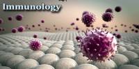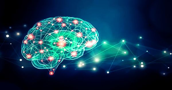
Cell signaling is the process by which a cell communicates with itself, other cells, and its surroundings. It is the process by which cells communicate with one another to coordinate various activities within a multicellular organism. It is also known as cell communication. Cell signaling is a fundamental property of all prokaryotic and eukaryotic cellular life. This communication is required for physiological processes such as growth, development, immune responses, and homeostasis to function properly.
Signaling typically consists of three components: the signal, the receptor, and the effector. It entails the transmission of signals, which are frequently in the form of molecules, from one cell to another. These signals can be classified into three main types:
- Autocrine Signaling: Cells release signaling molecules that bind to receptors on their own cell surface in autocrine signaling. This enables the cell to respond to its own signals, which is critical in processes like cell growth and differentiation.
- Paracrine Signaling: The release of signaling molecules into the extracellular fluid affects nearby cells via paracrine signaling. These signals have receptors in the target cells, and the effects are typically localized to a specific tissue or region.
- Endocrine Signaling: Signaling molecules (hormones) are released into the bloodstream and carried to distant target cells with the appropriate receptors in endocrine signaling. This type of signaling is essential for coordinating activities throughout the body.
In biology, signals are mostly chemical in nature, but can also be physical cues such as pressure, voltage, temperature or light. Chemical signals are molecules with the ability to bind and activate a specific receptor. These molecules, also referred as ligands, are chemically diverse including ions (e.g. Na+, K+, Ca++, etc.), lipids (e.g. steroid, prostaglandin), peptides (e.g. insulin, ACTH), carbohydrates, glycosylated proteins (proteoglycans), nucleic acids, etc.
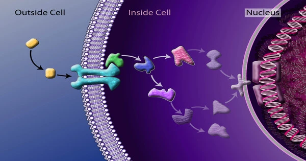
The process of cell signaling can be broken down into several key steps:
- Signal Reception: The target cell detects the signaling molecule by binding it to a specific receptor protein, usually located on the cell surface or within the cell.
- Transduction: When the signaling molecule binds to the receptor, it undergoes a conformational change, triggering a series of intracellular events known as signal transduction. This frequently involves a chain reaction of molecular events within the cell.
- Cellular Response: The signal transduction pathway results in a cellular response, which can include changes in gene expression, protein activity changes, or other cellular processes. The response is unique to the signal type and the target cell.
Peptide and lipid ligands are particularly important because most hormones fall into these chemical classes. Peptides are typically hydrophilic, polar molecules. As a result of their inability to diffuse freely across the plasma membrane’s bi-lipid layer, their action is mediated by a cell membrane bound receptor.
Liposoluble chemicals, on the other hand, such as steroid hormones, can diffuse passively across the plasma membrane and interact with intracellular receptors. Cell signaling can be autocrine, intracrine, juxtacrine, paracrine, or endocrine, and can occur over short or long distances.
When a chemical signal acts on the same cell that produced the signaling chemical, this is known as autocrine signaling. Intracrine signaling occurs when a chemical signal produced by a cell acts on receptors located in the same cell’s cytoplasm or nucleus. Juxtacrine signaling occurs between cells that are physically adjacent. Paracrine signaling occurs between cells that are close together. Endocrine interaction takes place between distant cells, with the chemical signal typically carried by blood.