The tissue optical clearing method, when combined with microscopic imaging technology, allows us to see various morphological details in the brain, allowing us to study the relationship between morphological structure and function as well as reveal pathological changes associated with disease occurrence.
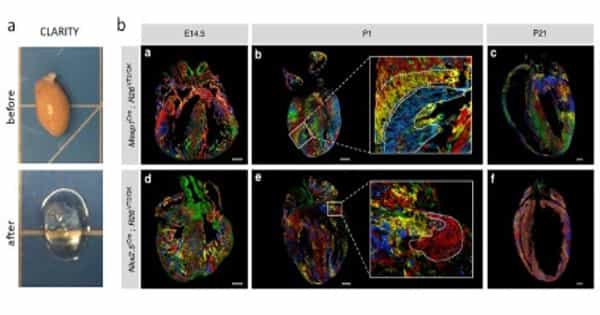
The ability to visualize cancerous tumors and metastatic tissue in three dimensions (3D) can aid clinicians in determining the exact type and stage of cancer, as well as the best treatment options. A Japanese research team tested the effectiveness of specialized hydrogels to obtain even clearer tissue for imaging. These hydrogels, which act as a 3D molecular network, can rapidly remove fats from tissues without losing their structure, which is a factor in tissue opacification. Contact lenses are among the biomedical devices that use the material.
When it comes to cancer, it’s important to be as clear as possible. The ability to visualize cancerous tumors and metastatic tissue in three dimensions (3D) can aid clinicians in determining the exact type and stage of cancer, as well as the best treatment options. A Japanese research team tested the effectiveness of specialized hydrogels to obtain even clearer tissue for imaging. These hydrogels, which act as a 3D molecular network, can rapidly remove fats from tissues without losing their structure, which is a factor in tissue opacification. Contact lenses are among the biomedical devices that use the material.
The ability to visualize cancerous tumors and metastatic tissue three-dimensionally (3D) can help clinicians diagnose the precise type and stage of cancer, while also informing the best treatment methods.
Cancer has been the leading cause of death in Japan since 1981 “Chie Kojima, an associate professor at Osaka Prefecture University’s Graduate School of Engineering’s Department of Applied Chemistry, was the study’s first author. “New treatment methods and diagnostic techniques are required. 3D fluorescence imaging is one such technique that could be crucial for understanding multicellular systems on an organ scale, as it provides more information than traditional 2D imaging. This could be useful for diagnosis and elucidating biological phenomena in personalized medicine.
This type of imaging entails labeling molecular machines like proteins so that they fluoresce in different colors depending on what they are. The glowing signals can be seen in a number of samples, ranging from whole organisms to single cells. However, most tissues are opaque, making it difficult to see these signals. The signals are visible in 2D imaging because the samples are thinly sliced, but the ability to visualize the entire system in 3D is lost.
Previously, researchers used the CLARITY method, which involves embedding tissues in polyacrylamide hydrogels. The fats in the tissues are removed, and the media’s refractive index is adjusted. The tagged glowing signals can be seen in 3D, but the cancerous tissue takes a month to clear, which Kojima believes is far too long for a patient waiting for a diagnosis. The tumor would have most likely spread by that time.
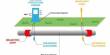
All tissue clearing techniques aim to make tissues or cell cultures more transparent in order to overcome their opacity, which prevents visible wavelengths of light from penetrating them under the microscope. It may be preferable to clear tissue through several chemical steps rather than going through the time-consuming process of cutting very thin sections of tissue, which can disrupt morphology. Clearing also allows for 3D microscopy, which produces images that are more reflective of biological structures than traditional 2D methods.
Typical tissue clearing methods include immersing the tissue in various solutions (such as an organic solvent or an aqueous solution with a high refractive index) or possibly embedding the tissue in a hydrogel before extracting tissue lipids with detergents. Depending on the type of tissue you use, different methods have a variety of advantages and disadvantages.
For practical applications, the optical clearing process time in the CLARITY method needs to be shortened,” Kojima said. The researchers used zwitterionic hydrogels, which have balanced charged molecules and hold the structure of tissue samples, to reduce this time. The team discovered that polymer hydrogels that mimic fatty molecules on the tissue appear to optically clear tumor tissues the fastest out of several zwitterionic hydrogel combinations. The hydrogels are highly osmotic, according to Kojima, which may aid in the removal of other fatty acids from the tissue.
“We were able to visualize blood vascular networks in murine brain tissues as well as metastatic tumor tissues in 3D using our system,” Kojima said. They could also visualize the tumor tissues faster than in previous attempts: what took a month before could now be done in a week with the new method.
The researchers are still looking into the technique and how it can be used to diagnose cancer in humans. “We’re trying to use our system for pathological diagnostics,” said Kojima. “Instead of thin slices, we expect to be able to diagnose a whole biopsy sample, which could prevent small cancers from being missed.”