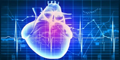
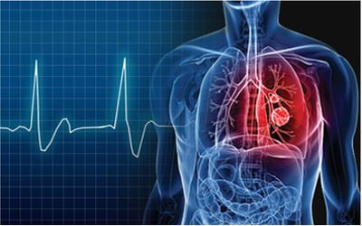
Electrophysiology is the study of the electrical properties of biological cells and tissues. It involves measurements of voltage change or electrical current flow on a wide variety of scales from single ion channel proteins, to whole tissues like the heart. This study is a test performed to assess your heart’s electrical system or activity and is used to diagnose abnormal heartbeats or arrhythmia. This helps keep blood flowing the way it should. This movement of the heart creates a heartbeat or heart rhythm.
In neuroscience, it includes measurements of the electrical activity of neurons, and particularly action potential activity. Neurons communicate using electrical and chemical signals. Electrophysiology techniques listen in on these signals by measuring electrical activity, allowing scientists to decode intercellular and intracellular messages.
Classical electrophysiologic techniques
Classical electrophysiology involves placing electrodes into various preparations of biologic tissue. The test is performed by inserting catheters and then wire electrodes, which measure electrical activity, through blood vessels that enter the heart. The principal types of electrodes are – (1) simple solid conductors, such as discs and needles (singles or arrays), (2) tracings on a printed circuit board, and (3) hollow tubes filled with an electrolyte, such as glass pipettes.
The principal preparations include – (1) living organisms, (2) excised tissue (acute or cultured), (3) dissociated cells from excised tissue (acute or cultured), (4) artificially grown cells or tissues, or (5) hybrids of the above.
If an electrode is small enough in diameter (on the order of microns), then the electrophysiologist may choose to insert the tip into a single cell. These techniques can answer systems-level questions, such as the role of a neuron in a neural circuit or behavior.
Many particular electrophysiological readings have specific names:
- Electrocardiography – for the heart,
- Electroencephalography – for the brain,
- Electrocorticography – from the cerebral cortex,
- Electromyography – for the muscles,
- Electrooculography – for the eyes,
- Electroretinography – for the retina,
- Electroantennography – for the olfactory receptors in arthropods.