Using tin mono-sulfide (SnS) nanosheets, researchers created the world’s thinnest X-ray detector. The new X-ray detector is less than 10 nanometers thick and highly sensitive, with a fast response time, and could one day lead to real-time imaging of cellular biology. Australian researchers created the thinnest X-ray detector ever made using tin mono-sulfide (SnS) nanosheets, potentially enabling real-time imaging of cellular biology.
X-ray detectors, like medical imaging or Geiger counters, are tools that allow radiation energy to be recognized visually or electronically. SnS has already demonstrated great promise in photovoltaics, field-effect transistors, and catalysis.
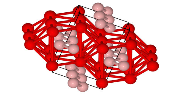
Members of the ARC Centre of Excellence in Excitation Science at Monash University and RMIT University have now demonstrated that SnS nanosheets are also excellent candidates for use as soft X-ray detectors. Their findings, published in the journal Advanced Functional Materials, show that SnS nanosheets have high photon absorption coefficients, allowing them to be used in the fabrication of ultrathin soft X-ray detectors with high sensitivity and fast response times.
These materials were discovered to be even more sensitive than another emerging candidate (metal halide perovskites), with a faster response time than established detectors and the ability to be tuned for sensitivity across the soft X-ray range.
The SnS nanosheets respond extremely quickly, within milliseconds. You can scan something and instantly get an image of it. The time resolution is determined by the sensing time. Using X-rays, you can see proteins and cells evolving and moving, rather than just a static image.
Professor Jacek Jasieniak
The team’s SnS X-ray detectors are less than 10 nanometres thick. To put things into perspective, a sheet of paper is approximately 100,000 nanometres thick, and your fingernails grow at a rate of one nanometre per second. Previously, the thinnest X-ray detectors were between 20 and 50 nanometres thick.
Although much work remains to be done to fully exploit the potential of the SnS X-ray detectors, the senior author of the paper, Professor Jacek Jasieniak of Monash’s Department of Materials Science and Engineering, believes that this could one day lead to real-time imaging of cellular processes.
“The SnS nanosheets respond extremely quickly, within milliseconds,” he explained.
“You can scan something and instantly get an image of it. The time resolution is determined by the sensing time. In theory, because of the high sensitivity and time resolution, you might be able to see things in real-time. You might be able to use this to observe how cells interact. Using X-rays, you can see proteins and cells evolving and moving, rather than just a static image.”
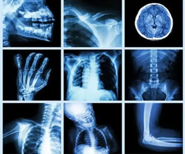
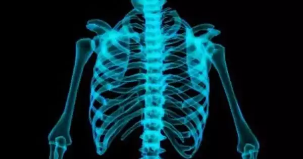
What is the significance of such sensitive and responsive detectors? X-rays are broadly classified into two types: Hospitals use ‘hard’ X-rays to scan the body for broken bones and other illnesses.
Soft X-rays, which have lower photon energy and can be used to study wet proteins and living cells, are a less well-known but equally important component of cellular biology. Some of these measurements are carried out in the ‘water window,’ a region of the electromagnetic spectrum where water is transparent to soft X-rays.
Soft X-ray detection can be performed using a Synchrotron, a particle accelerator similar to Switzerland’s Large Hadron Collider, but access to this type of massively expensive infrastructure is difficult to secure. Recent advances in non-synchrotron soft X-ray laser sources may enable the development of lower-cost, portable detection systems, providing a viable alternative to Synchrotrons for researchers worldwide.
However, in order for this approach to work, we will need soft X-ray detector materials that are highly sensitive to low energy X-rays, have a high spatial resolution, and are inexpensive. Some existing soft X-ray detectors employ an indirect mechanism that converts ionizing radiation into visible photons. This approach allows for multiple energy ranges and frame rates to be studied but is difficult to prepare and offers a limited resolution.
Because the detector material can be thinner in direct detection methods, they are easier to prepare and provide higher resolutions than indirect methods. A high X-ray absorption coefficient is required for good candidate materials, which is calculated using the atomic number of the absorbing atoms, X-ray incident energy, density, and atomic mass of an atom.
Soft X-rays are more strongly absorbed in thin materials than hard X-rays due to their high atomic mass and low energy. Although nanocrystal films and ferromagnetic flakes have shown promise as soft X-ray detectors, they are not well suited for use in the water region. This is where SnS nanosheets come into play.
Dr. Nasir Mahmood of RMIT University, one of the lead authors, stated that the sensitivity and efficiency of SnS nanosheets are highly dependent on their thickness and lateral dimensions, which are difficult to control using traditional fabrication methods.
Using a liquid metal-based exfoliation method, the researchers were able to create high-quality, large-area sheets with a controlled thickness that can detect soft X-ray photons in the water region. Their sensitivity can be increased further by stacking the ultrathin layers.
They provide significant improvements in sensitivity and response time over existing direct soft X-ray detectors. The researchers hope that their discoveries will pave the way for the development of next-generation, highly sensitive X-ray detectors based on ultrathin materials.
Dr. Babar Shabbir of Monash University’s Department of Materials Science and Engineering, the study’s first author, stated: “In the long run, we’ll need to test a multi-pixel device in order to commercialize this. We don’t have an imaging system at this time. However, this provides us with a knowledge platform as well as a prototype.”