Papilledema
Definition
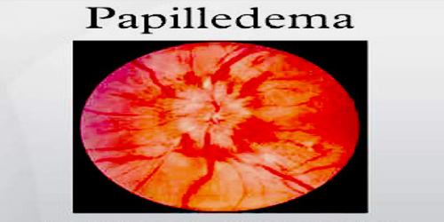
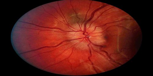
Papilledema is the swelling of the optic disc caused by increased intracranial pressure. The optic nerve head, also called the optic disk or papilla, is the area where the optic nerve (the nerve that carries messages from the eye to the brain) enters the eyeball. The swelling is usually bilateral and can occur over a period of hours to weeks. Unilateral presentation is extremely rare. Papilledema is mostly seen as a symptom resulting from another pathophysiological process.
The optic nerve is anatomically continuous with the brain through the subarachnoid space. Any increase in pressure in the cranium thereby is being transmitted to the optic nerve. The anterior end of the optic nerve discontinues at the eye, thereby causing protrusion of the optic nerve on the area.
The causes of papilledema include cerebral edema (swelling of the brain, as from encephalitis or trauma), tumors and other lesions that occupy space within the skull, increased production of cerebrospinal fluid (CSF), decreased resorption of CSF (due to venous sinus thrombosis, meningitis, or subarachnoid hemorrhage), obstruction of the ventricular system within the brain, hydrocephalus, craniosynostosis (premature closure of the sutures of the skull), and a condition called pseudotumor cerebri.
Causes, Sign and Symptoms of Papilledema
Optic nerve swelling can happen when CSF builds up where your optic nerve and the central retinal vein travel between our brain and our eye nerve. This area is known as the subarachnoid space. When pressure pushes on the nerve and vein, blood and fluid can’t leave the eye at a normal rate, causing papilledema. Brain swelling can be caused by a number of injuries and conditions, including:
- traumatic injury to our head
- not having enough red blood cells or hemoglobin (anemia)
- CSF buildup in our brain (hydrocephalus)
- brain bleeding (hemorrhage)
- brain inflammation (encephalitis)
- brain tissue inflammation (meningitis)
- high blood pressure (hypertension)
- collection of infected pus in the brain (abscess)
- brain tumor
Sometimes, brain pressure builds up for no apparent reason and this is known as idiopathic intracranial hypertension.
In the early stages, papilledema may be asymptomatic or present with a headache. It can progress to enlargement of the blind spot, blurring of vision, visual obscurations. Ultimately, total loss of vision can occur. The signs of papilledema that are seen using an ophthalmoscope include:
- venous engorgement (usually the first signs)
- loss of venous pulsation
- hemorrhages over and/or adjacent to the optic disc
- blurring of optic margins
- elevation of the optic disc
- Paton’s lines (radial retinal lines cascading from the optic disc)
The eyes are examined using an opthalmoscope and may reveal loss of venous pulsation, venous engorgement, hemorrhages, optic disc elevation and radial retinal lines termed as Paton’s lines.
Diagnosis and Treatments of Papilledema
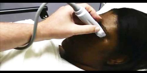
The diagnosis of papilledema relies on the use of slit lamp examination and opthalmoscopy showing a characteristic of the optic nerve disc. The diagnostic tests are done when new onset headache is noted with possible increased intracranial pressure. Aside from the optic nerve diagnosis, a biopsy can be made to detect whether the tumor is benign or malignant whenever it is present. Papilledema is usually differentiated from other conditions involving the eyes such as papillitis, optic neuritis, glaucoma, and retinal detachment.
Doctor may prescribe medications to reduce swelling. Corticosteroids, such as prednisone (Deltasone), dexamethasone (Ozurdex), and hydrocortisone (Cortef), can all be used to keep swelling down in our brain. These medications may be injected or taken by mouth.
If high blood pressure is causing papilledema, doctor may prescribe medications to keep patients blood pressure under control. Common medications for high blood pressure include:
- Diuretics: bumetanide (Bumex) and chlorothiazide (Diuril)
- Beta blockers: atenolol (Tenormin) and esmilol (Brevibloc)
- ACE inhibitors: captopril and moexipril
Other treatments include repeated lumbar punctures to remove excess spinal fluid in the cranium. The removal of potentially causative medicines including tetracyclines and vitamin A analogues may help decrease ICP; however, this is only necessary if the medication is truly felt to contribute to the ICP increase.
Papillitis is a condition of optic neuritis. It is called intraocular optic neuritis wherein there is inflammation of the optic nerve head. Papilledema on the other hand, is the bulging or swelling of the optic disc. Papillitis involves significant loss in the visual field and pain on the globe of the eyes, which is not present in papilledema.
Reference: